

Applications
3D & 2D Cell Culture and Imaging
- Culture and visualize three-dimensional (3D) cell aggregates and two-dimensional (2D) monolayers
- Ideal for spheroids, organoids, embryoid bodies, and stem cells
- Enables long-term cell growth and microscopy readouts
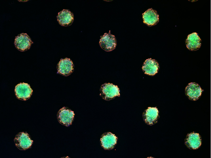
Scaffold-Free 3D Cell Models
- Supports the self-organization of 3D cell structures
- Maintains native cell behavior for accurate experimental results
High-Resolution Fluorescence & Live Cell Imaging
- Compatible with fluorescence microscopy, allowing high-resolution imaging of live and fixed cells
- Supports immunofluorescence staining for in-depth subcellular analysis
- Designed for long-term live cell imaging experiments
Other Multi-Cell Patterned Formats
- Pattern sizes: also available with 500 µm diameter and 1000 µm pitch (#83613)
- Labware format: also available in the µ-Slide 8 Wellhigh
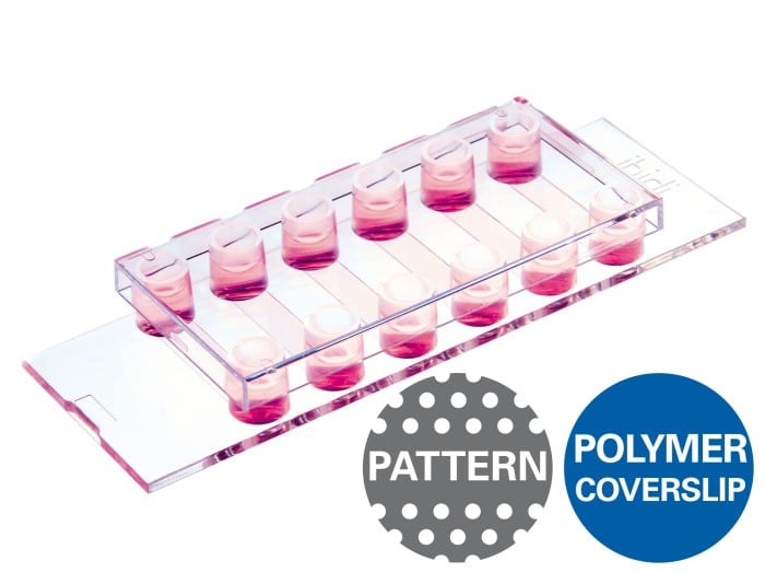
Technical Features:
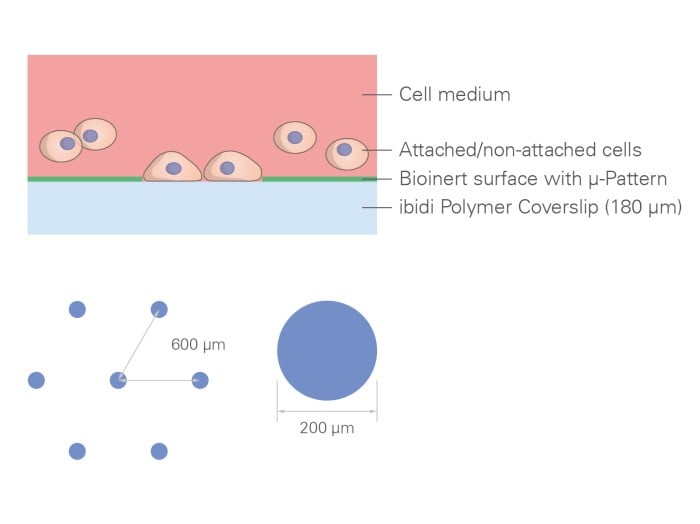
- Precision µ-Patterning – The µ-Slide features an ibiTreat micropatterned surface enclosed by an advanced ibidi Bioinert surface, ensuring selective cell adhesion.
- Exclusive Bioinert Surface – Outperforms standard ultra-low attachment (ULA) surfaces:
- No unwanted cell or protein adhesion
- Long-term stability for days or weeks
- Biologically inert, preserving cell viability and behavior
- Superior Imaging Compatibility – The Bioinert surface is applied to the ibidi Polymer Coverslip, delivering unmatched optical clarity for high-resolution microscopy.
- Non-Fluorescent, Phase Contrast Compatible – Patterns remain non-fluorescent but slightly visible under phase contrast, facilitating easy alignment and analysis.
- Compatibility - Fully biocompatible materials, compatible with different protein and peptide coatings as well as with staining and fixation solutions
- Different Slide Formats - Available on either the µ-Slide 8 Well high or µ-Slide VI 0.4