Athena Zebrafish Image Analysis Software

SKU: - UNSPSC: - Manufacturer Part No: - Pack Size: -
  • Quick & simple user interface

  • Designed for non-specialists in image analysis

  • Compatible for images from commercial microscopes

  • The fastest & easiest Zebrafish image-based screening tool available

  • Novel Artificial Intelligence-based algorithms for fish & organ identification

  • No user intervention is required to readily detect fish & internal organs in brightfield

Special Requirements?

Build out your order then request a quote from your cart.

Clear
See Variant info VAT
Ex. Inc.

Product succesfully added to cart. View Cart

If you are looking for a quote, you can request this here.

IDEA Bio-Medical is proud to present Athena Zebrafish – the next generation in Zebrafish assays. Our new, unique dedicated analysis software for automated analysis of Zebrafish microscopy images offers simple and quick quantification of fluorescence, measurement of morphological changes & other phenotypic features in Zebrafish larvae in a high throughput format.

Athena Zebrafish is suited for a broad range of researchers and accepts multiple image format types output from nearly all microscope manufacturers.

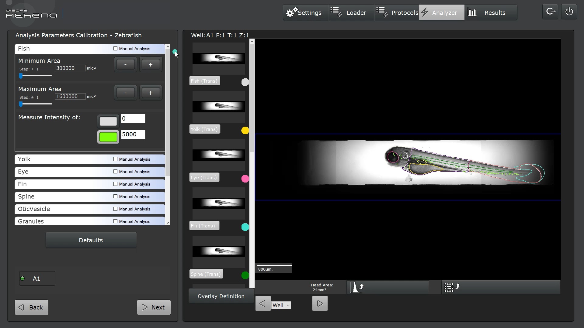
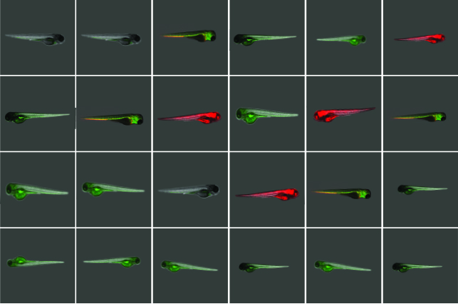
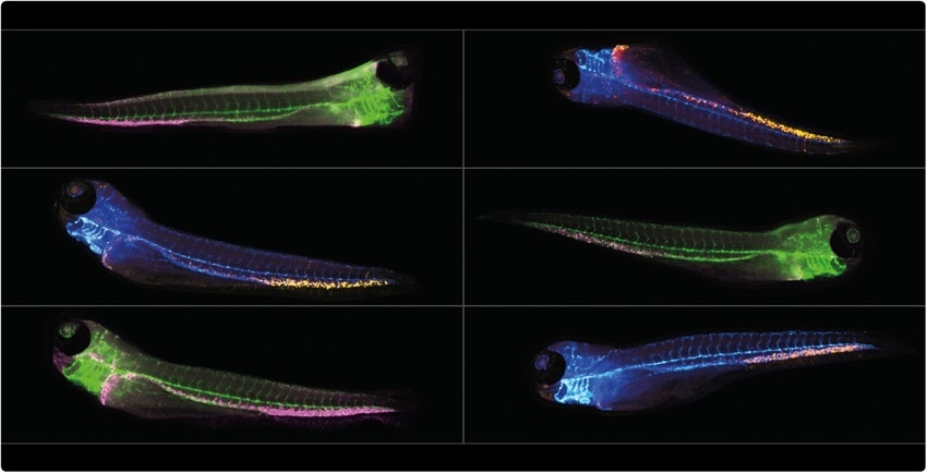
The software permits parameter-free zebrafish analysis using simple bright-field images. It automatically detects zebrafish embryos and larvae up to 5 days old (dpf), extracting the fish contour and much of its internal anatomy: yolk sac, eye, notocord, and more, along with body regions of the head, trunk, and tail.

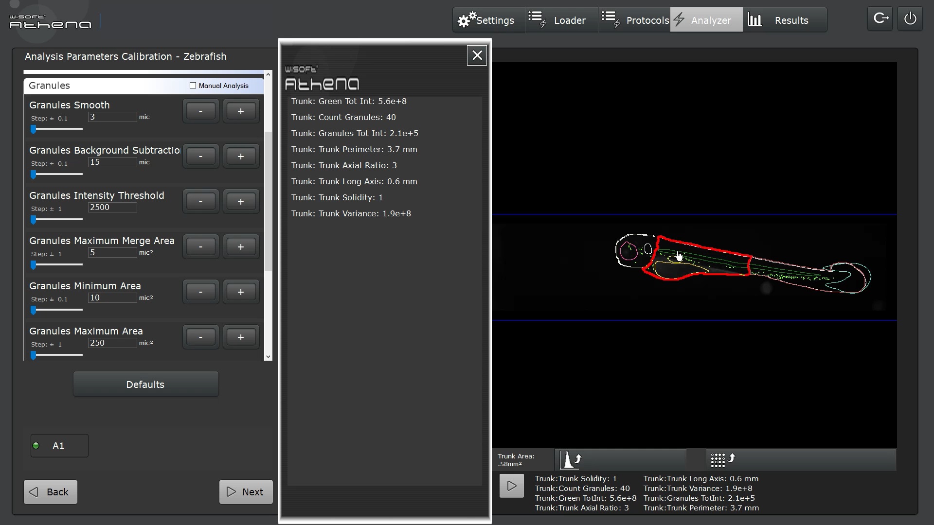
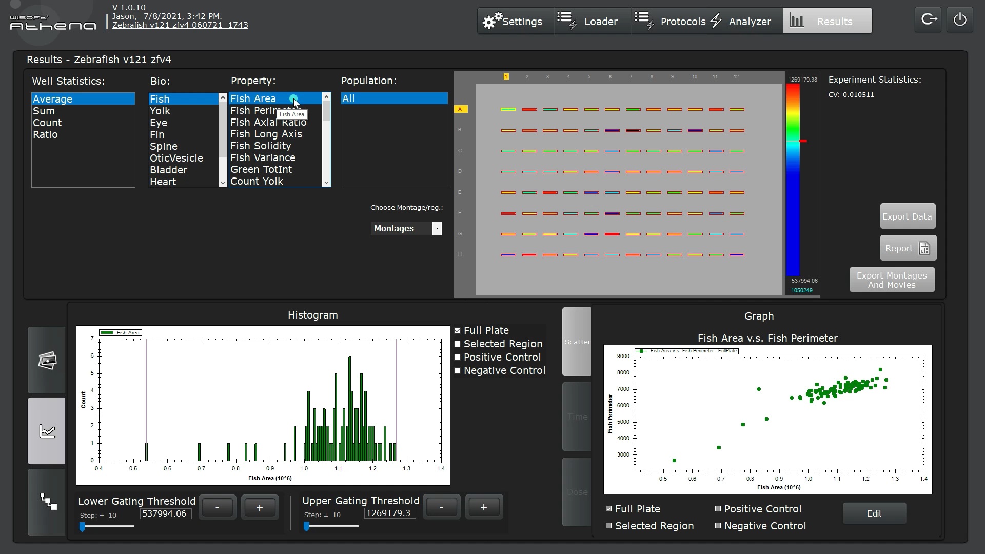
For each of these objects, the software measures the morphology (area, length, and shape) and can detect fluorescence in associated color channels. Both fluorescence intensity and spot/structure detection within specific anatomy are supported.

Zebrafish (Danio rerio) are an attractive model organism for the study of human disease pathology because of their optical transparency and genetic tractability. They serve as a great alternative to mammalian screening due to cost, throughput and reduced ethical concerns.

Automated analysis of Zebrafish imposes unique demands due to the versatility of organs and features needed to be detected.

To increase throughput, robustness and permit unbiased image analysis of zebrafish, IDEA Bio-Medical developed a novel AI-based software to automatically  detect fish contour and internal anatomy in brightfield images.  By identifying these hard to detect structures, our software maintains the anatomical context of associated fluorescence signals to enable true high-content imaging in Zebrafish.  Thusly, zebrafish morphology along with region-specific spot/cell counting and fluorescence intensity measurement are readily quantified.

  • Quick & simple user interface

  • Designed for non-specialists in image analysis

  • Compatible for images from commercial microscopes

  • The fastest & easiest Zebrafish image-based screening tool available

  • Novel Artificial Intelligence- based algorithms for fish & organ identification

  • No user intervention required to readily detect fish & internal organs in brightfield

  • Combine with fluorescent labeling for structural co-analysis

  • Ensure proper fish orientation in post-analysis with customizable, software-based selection

  • Software  suited for researchers who only image several fish per week, as well as researchers imaging hundreds & thousands of fish in multi-well plates for large scale screens.

  • Software accepts any microscopy image, supporting multiple image formats from manual microscopes or automated imaging systems from vendors such as Leica, Zeiss, Nikon and more.

  • Unbeatable throughput: analyze hundreds of Zebrafish larvae within minutes

  • Analyzes  Label-free or fluorescently tagged fish and internal organelles

  • Analyze your Fish larva from head to tail with supported image analysis from single plane, Z stack and projection images

Computer requirements

  • Hard Disk space available: 6GB minimum.
  • Minimum 8GB RAM, preferable 16GB.
  • At least 2 cores, preferable 8.
  • Monitor screen: To ensure proper software visibility, screen resolution should be set to at least 1920×1080 and the text scaling should be set to 100%.

Image formats

Athena Zebrafish supports loading a broad range of image formats either with or without the associated Metadata. Images must be either 8-bit or 16-bit grayscale images, as RGB color images are not supported.

Without metadata, supported image formats are *.tif, *.tiff, *.png, *.bmp, *.jpg, *.jpeg. For these formats, users can manually define pixel size and the software can identify associated color channels present in separate image files. To group the color channels together, identifying text in the file name of each image should be present e.g., Brightfield_01.tif,  Green_01.tif, Red_01.tif. Multi-page *.tif image stacks are accepted when each page corresponds either to a Z- or T-stack (time-lapse). Each Z- or T-slice is analyzed separately and 3D rendering is not supported.

When loading OME-TIFF and proprietary formats (*.czi, *.nd2, etc), Athena can automatically extract the following associated metadata:

  1. Dimensions of each image: well, in well field, wavelength, time index, Z-stack index. 
  2. Pixel size
  3. Plate info
  4. Objective

Image Characteristics

Athena Zebrafish has four requirements to visualize zebrafish embryos or larvae visualized in a micrograph image:

  1. Analysis of Zebrafish larvae and embryos is supported, up to 5 DPF (5 days post fertilization).
  2. The zebrafish animal must be imaged using transmitted light for contour and anatomy detection. Whole-body fluorescence images are not supported.
  3. The transmitted light channel must have a bright (white) background. Best results are obtained for zebrafish imaged with standard brightfield illumination rather than contrast enhanced transmitted light methodologies (i.e. phase contrast, DIC).  
  4. The whole animal must be visible within the image, from head to tail. Images visualizing only specific regions, such as the heart or tailfin, are not yet supported.
Variant
Pack/Size
Ordering
Price
Qty
Zebrafish-software-opening-screen
500 Images
500 / Images

SKU: IDA-ATHENA500

POA
Zebrafish-software-opening-screen-1
1000 Images
1000 / Images

SKU: IDA-ATHENA1000

POA
Zebrafish-software-opening-screen-2
5000 Images
5000 / Images

SKU: IDA-ATHENA5000

POA
Zebrafish-software-opening-screen-3
10000 Images
10000 / Images

SKU: IDA-ATHENA10000

POA
Zebrafish-software-opening-screen-4
25000 Images
25000 / Images

SKU: IDA-ATHENA25000

POA
Zebrafish-software-opening-screen-5
50000 Images
50000 / Images

SKU: IDA-ATHENA50000

POA

Newsletter Sign Up

  • Hidden
  • Hidden
  • Hidden
    DD slash MM slash YYYY
  • Hidden
  • Hidden
  • Hidden

Need a new Thermal Cycler?

We’re offering some amazing deals when you trade in your exisitng thermal cycler for recycling. Just contact us to find out more.

Call 01698 338844

Athena Zebrafish Image Analysis Software

Web Scientific has now joined our sister site Thistle Scientific!

We’re the same great team with the same commitment to quality, but now you can browse a larger range with much greater ease and selling points to say why this is a benefit to you.